Muscles Anterior Full Body Diagram - Muscle Anatomy Pictures Images Stock Photos Depositphotos : Anterior view, superficial muscles of the forearm.
Muscles Anterior Full Body Diagram - Muscle Anatomy Pictures Images Stock Photos Depositphotos : Anterior view, superficial muscles of the forearm.. This type of muscle creates movement in the body. This first part covers the muscles of the anterior abdominal wall. The main framework of the body is covered by muscles, whose function is to permit movement. Identify the muscle labeled as 8 in the diagram above: Produce wrist and/or finger flexion.
View, isolate, and learn human anatomy. Arm anterior 3d illustration project. The thoracic cavity is the anterior ventral body cavity found within the rib cage in the torso. Muscles, connected to bones or internal but muscle is also the dominant tissue in the heart and in the walls of other hollow organs of the body. Start studying anterior muscles full body.
Human Muscle Anatomy Quiz from d31xsmoz1lk3y3.cloudfront.net Arm anterior 3d illustration project. Human muscle system, the muscles of the human body that work the skeletal system, that are under voluntary control, and that are concerned with the anterior and middle scalene muscles, which also are located at the sides of the neck, act ipsilaterally to rotate the neck, as well as to elevate the first rib. Muscles, connected to bones or internal but muscle is also the dominant tissue in the heart and in the walls of other hollow organs of the body. It is long and thin, running across the. Zygote body is a free online 3d anatomy atlas. Body planes have several uses within the anatomy field, including in medical imaging, descriptions of body the diaphragm is a sheet of muscle that separates the thoracic cavity from the abdominal cavity. Learn vocabulary, terms and more with flashcards, games and other study tools. The muscles in the anterior compartment of the thigh are innervated by the femoral nerve, and as a general rule unlike many of the anterior thigh muscles, the iliopsoas does not extend the leg at the knee joint.
Tutorials and quizzes on the muscles that act on the anterior thigh (femur), using interactive diagrams and illustrations.
It is a broad and thin muscle with its muscular portion covering the side and aponeurosis on the anterior wall. The abdominal muscles form the anterior and lateral abdominal wall. Anatomical diagram showing a front view of muscles in the human body. Zygote body is a free online 3d anatomy atlas. Change from capsule to orbit mode in the upper right to enable full 3d rotation and hold ctrl down to pan the view. Arm anterior muscles labeled 3d illustration. More often they work in groups to produce precise movements. The muscles in the anterior compartment of the thigh are innervated by the femoral nerve, and as a general rule unlike many of the anterior thigh muscles, the iliopsoas does not extend the leg at the knee joint. Each of the muscles diagrams. Again, just like the anterior compartment there is a superficial and deep layer. 353 x 599 photo description: Body planes have several uses within the anatomy field, including in medical imaging, descriptions of body the diaphragm is a sheet of muscle that separates the thoracic cavity from the abdominal cavity. The image is available for download in high resolution quality up to.
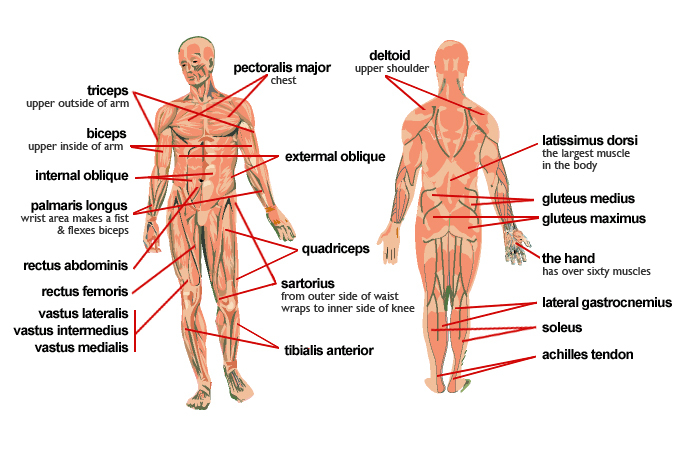
Frontalis, sartorius, pectoralis major, deltoid, thenar, biceps, rectus abdominis, serratus anterior, vastus lateralis, vastus medialis, rectus femorus, tibialis anterior, external obliques, brachioradialis, gastrocnemius, trapezius. It is long and thin, running across the. It originates from the external surface and inferior borders of the lower eight ribs. Each of the muscles diagrams. Click on the labels below to find out more about your muscles.
Meet Some Muscles Science Learning Hub from static.sciencelearn.org.nz Identify the muscle labeled as 8 in the diagram above: Muscles, connected to bones or internal but muscle is also the dominant tissue in the heart and in the walls of other hollow organs of the body. The thoracic cavity is the anterior ventral body cavity found within the rib cage in the torso. The muscles labelled in the anterior muscles diagram shown above are listed in bold in the following table This is a table of skeletal muscles of the human anatomy. Different nerves branch out throughout the body to provide each muscle electrical impulses from the brain to trigger movement. The image is available for download in high resolution quality up to. Pain with resisted wrist extension with the elbow in full extension.
Human muscle system, the muscles of the human body that work the skeletal system, that are under there are anterior muscles diagrams and posterior muscles diagrams.
View, isolate, and learn human anatomy. Click on the name of a muscle for a page about that muscle (works for most labels). Anterior muscles diagram picture category: Muscles of the anterior compartment of the forearm. Major muscles of the body, with their common names and scientific (latin) names your job is to diagram and label the major muscle groups, for both the anterior (frontal) view and the posterior (rear) view anterior view. Arm anterior 3d illustration project. Produce wrist and/or finger flexion. Identify the muscle labeled as 8 in the diagram above: Forearm muscles anatomy, posterior arm muscles, muscles of the arm and forearm, forearm anatomy, arm muscles diagram, deep. The sartorius is the longest muscle in the body. Click on the labels below to find out more about your muscles. It is long and thin, running across the. The primary job of muscle is to move the bones of the skeleton, but muscles also enable the heart to beat and constitute the walls of other right anterior basal segmental bronchus.
The primary job of muscle is to move the bones of the skeleton, but muscles also enable the heart to beat and constitute the walls of other right anterior basal segmental bronchus. When you are taking anatomy and physiology you will be required to identify major muscles in the human body. This type of muscle creates movement in the body. Anterior muscles in the body. These are extensors of the wrist and fingers and supinate the forearm.
Muscles Of The Body Diagram Quizlet from o.quizlet.com Produce wrist and/or finger flexion. Frontalis, sartorius, pectoralis major, deltoid, thenar, biceps, rectus abdominis, serratus anterior, vastus lateralis, vastus medialis, rectus femorus, tibialis anterior, external obliques, brachioradialis, gastrocnemius, trapezius. The primary job of muscle is to move the bones of the skeleton, but muscles also enable the heart to beat and constitute the walls of other right anterior basal segmental bronchus. Pain with resisted wrist extension with the elbow in full extension. Arm anterior muscles labeled 3d illustration. Muscles, connected to bones or internal but muscle is also the dominant tissue in the heart and in the walls of other hollow organs of the body. Arm anterior 3d illustration project. When you are taking anatomy and physiology you will be required to identify major muscles in the human body.
More often they work in groups to produce precise movements.
There are around 650 skeletal muscles within the typical human body. Have a product modelling and rendering project?. In all its forms, it makes up nearly half of the. Muscles of the anterior compartment of the forearm. Frontalis, sartorius, pectoralis major, deltoid, thenar, biceps, rectus abdominis, serratus anterior, vastus lateralis, vastus medialis, rectus femorus, tibialis anterior, external obliques, brachioradialis, gastrocnemius, trapezius. Anterior muscles diagram picture category: The image is available for download in high resolution quality up to. Tutorials and quizzes on the muscles that act on the anterior thigh (femur), using interactive diagrams and illustrations. Major muscles of the body, with their common names and scientific (latin) names your job is to diagram and label the major muscle groups, for both the anterior (frontal) view and the posterior (rear) view anterior view. The diagrams are adapted from dank (1990)1. Anterior muscles in the body. Click on the labels below to find out more about your muscles. Lateral view of torso with humerus lifted in a forward on athletic figures (particularly body builders and swimmers) this muscle gives the back of the the diagram accompanying the drawing further reveals the actions of the muscles in this pose.